RSS

Cell Membrane Structure and Function

phospholipid

plasma cell membrane

cell membrane coloring

cell membrane analogy

cell membrane animation
Membranes are vital  because they separate the cell from the outside world.  They also separate compartments inside the cell to protect important processes and events.

Cellular membranes have diverse functions in the different regions and organelles of a cell. However, at the electron microscopic level, they share a common structure following routine preparative steps. The above figure shows the typical "Unit" membrane which resembles a railroad track with two dense lines separated by a clear space.  This figure actually shows two adjacent plasma membranes, both of which have the "unit membrane" structure.  You can best see protein distribution via a technique called freeze fracture/freeze etch.  The freeze-fracture/freeze etch technique starts with rapid freezing of a cell. Then the frozen cells are cleaved along a fracture plane. This fracture plane is inbetween the leaflets of the lipid bilayer , as shown by this cartoon. The two fractured sections are then coated with heavy metal (etched) and a replica is made of their surfaces. This replica is then viewed in an electron microscope. One sees homogeneous regions where there was only the exposed lipid leaflet.
In certain areas of the cell, one also sees protrusions or bumps. These are colored red in the cartoon. Sometimes one can see structure within the bumps themselves. These are the transmembrane proteins.

The following illustration will show you a freeze-fracture/freeze etch view. The organization or structure of the transmembrane proteins can often be visualized.

  • Digg
  • Del.icio.us
  • StumbleUpon
  • Reddit
  • RSS

Osmosis Practical with Eggs

osmosis experiment with eggs

osmosis experiment with eggs hypothesis

egg experiment

osmosis experiment with eggs conclusion
Osmosis is the process by which water enters our tissues. All of our cells are surrounded by a membrane that selectively allows in anything the cell needs but prevents unwanted molecules from entering.

This works because the membrane contains lots of tiny holes that will let anything smaller through but obviously stop big molecules from entering the cell. Water is small enough to get through and enters by a process called diffusion - this means that it will travel from an area with a high water concentration to an area with a lower concentration until it is balanced out with both areas having the same concentration (known as isotonic).

An egg also has a membrane surrounding it so we can use it to represent a cell and see how osmosis works.

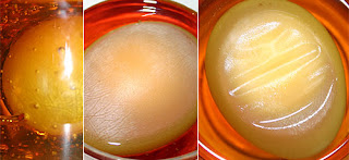
The first stage of the experiment was to soak the egg in vinegar. You will have seen that the shell completely disappears. Vinegar in fact contains acetic acid and this reacts with the calcium carbonate making up the shell of the egg. This reaction gives off carbon dioxide so if you were paying attention you will have seen bubbles coming off the egg as soon as it was added to the vinegar.

The membrane around the egg also becomes very rubbery during this soaking in vinegar. This occurs because the acid denatures the protein making up the egg white (albumin). Denaturation of proteins can occur through exposure to acids, bases or high temperature. This is what happens when you cook an egg- the clear egg white turns opaque and white as the protein chains become tangled due to the heat.

If you soak a cooked egg in vinegar it also becomes very rubbery - so much so that it will bounce when dropped from a low height! It's not a good idea to try this will an uncooked egg as inside the rubbery membrane the yolk and white are still runny and it will make a big mess if split!

So after soaking in vinegar you should have also noticed that the egg increases a little in size. This is because the water in the vinegar can enter the egg through the membrane, moving from the higher water concentration in vinegar to the lower concentration in the egg.

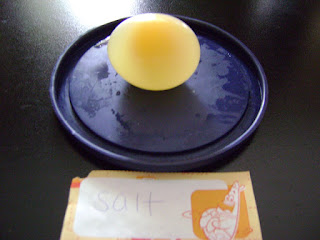
If you now put the egg in water, it will grow in size a lot more due to the much larger water concentration gradient across the membrane. If you add food coloring to the water you can see the process of osmosis in action as the colored water passes into the egg. Water is known as hypotonic, ie. very dilute and contains more water than the egg.

If instead you place the vinegar soaked egg in corn syrup you will see the opposite. There is a much higher water concentration in the egg than in the syrup so water will pass in the opposite direction. This means that the egg will shrink in size. The corn syrup is a hypertonic liquid, ie. very concentrated with not much water compared to the egg.

  • Digg
  • Del.icio.us
  • StumbleUpon
  • Reddit
  • RSS

Osmosis Potato Experiment

osmosis potato experiment

osmosis experiment lab report

osmosis experiment results
 osmosis potato experiment
Fill both of the dishes with water and add about two tablespoons of salt to one of the dishes. Using the knife have a parent cut the potato in half lengthwise. Then Place each piece flat side down in to one of the plates of water. Now simply let the two potato pieces soak in the water for a few hours. After this time has passed flip each potato over and look for differences. When looking at the potato pieces you can clearly see a difference between the two. Lets take a closer look at each of the potato pieces! This potato slice is the one that has been soaking in freshwater. Not much of a difference here, only that the potato is a little more rigid then before. This is because there is the more salt and other dissolved chemicals within the potato then the surrounding water. This means that the water will move into the potato.  This potato slice is the one that has been soaking in saltwater. This potato pieces looks substantially different from the original and the other slice. It seems to have wilted, gotten very soft and flexible. Why did that happen?

It has to do with a process called osmosis. The potato is made up of tiny, living units called cells. Each cell is surrounded by a cell membrane which acts much as your skin does. It keeps the cells parts inside and keeps other things outside, protecting the cell.

While this membrane stops most things, water can pass through it. The water tends to move towards higher concentrations of dissolved chemicals. That means that if the water outside the cell is saltier than the water inside, water will move from the inside of the cell to the outside. As the water left the cell it was much like letting the air out of a balloon. As more and more of the cells lost water, the slice of potato became soft and flexible.

  • Digg
  • Del.icio.us
  • StumbleUpon
  • Reddit
  • RSS

Onion Epidermis, Slides, Cells

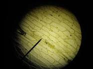
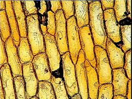
onion epidermis is composed of plant cells. These plant cells are arranged in order, because their cell walls obligations to each other. The epidermis appears to be of color. Many nuclei were found in the epidermis.


 onion epidermis,onion epidermis under microscope, onion microscope, image of cells, plasmolysis
 onion epidermis,onion epidermis under microscope, onion microscope, image of cells, plasmolysis


onion epidermis,onion epidermis under microscope, onion microscope, image of cells, plasmolysis

  • Digg
  • Del.icio.us
  • StumbleUpon
  • Reddit
  • RSS

Hydrilla: Hydrilla Leaf Cells

Hydrilla verticillata leaves a large amount of plant cells clearly defined. The leaf has a dark bar in the center. It's edges are uneven with strong bit "thorns" at the end. The leaf surface is similar to onion epidermis, but the nuclei of cells in plant leaves are barely visible anyway. The cells appear to be aligned in columns that all go from one end of the blade at its end and tapering approaches. Some cells appear slightly darker than appears to have removed most of the blue stain that most of the others.

 leaf cells, hydrilla leaf, hydrilla leaf cell, hydrilla cells, hydrilla, hydrilla plant, hydrilla verticillata, cabomba, hydrilla scientific name, pictures of hydrilla   


leaf cells, hydrilla leaf, hydrilla leaf cell, hydrilla cells, hydrilla, hydrilla plant, hydrilla verticillata, cabomba, hydrilla scientific name, pictures of hydrilla   

  • Digg
  • Del.icio.us
  • StumbleUpon
  • Reddit
  • RSS